


Stage
01
Inquiry
Tell us about your condition. Speak with our medical advisors — no obligation.
A combined optic-nerve and hormonal condition treated with targeted stem cell delivery. See visual, developmental and endocrine improvements documented in past septo-optic dysplasia patients. 81% reported quality-of-life improvement. 83% satisfied with the treatment outcome.
Published
Since 2007, we have been developing comprehensive stem cell treatment protocols for Septo-Optic Dysplasia (SOD) to overcome the limitations of conventional therapies. Septo-optic dysplasia (SOD) – also known as de Morsier syndrome – is a subtype of Optic Nerve Hypoplasia (ONH) and results from underdevelopment of the optic nerve, pituitary gland dysfunction, and absence of the septum pellucidum, which is a midline area of the brain. SOD arises from defects during the embryological development of infants and studies show that ONH maybe be related to gene defects as well as embryo exposure to infections. Read on to see if Septo-optic dysplasia Stem Cell Treatment might be right for your loved one.
Since 2007, we have been developing comprehensive stem cell treatment protocols for Septo-Optic Dysplasia (SOD) to overcome the limitations of conventional therapies. In our protocols, stem cells are combined with specialized therapies for SOD that not only focus on helping the patient to cope with their symptoms, but also treat the root cause of the condition by promoting the healing of the optic nerve and other affected brain structures. We believe that our comprehensive treatment approach for SOD gives our patients the best chances for vision improvement, allowing for a better quality of life.
Septo-Optic Dysplasia (SOD) is a rare congenital condition that involves underdevelopment of the optic nerves, often leading to significant vision impairment or blindness. The optic nerve, responsible for transmitting visual information from the eye to the brain, can be underdeveloped or affected by various factors in SOD, contributing to the challenges patients face.
While the exact causes of optic nerve hypoplasia in SOD are not always clear, it is often associated with disruptions during early brain and ocular development. Other general factors that can exacerbate or mimic optic nerve damage include:
Emerging research into stem cell therapy offers a promising avenue for addressing optic nerve damage in conditions like Septo-Optic Dysplasia. Stem cells have the potential to regenerate damaged tissues and improve the function of underdeveloped optic nerves. Although still in experimental stages, advancements in stem cell treatment may revolutionize care for SOD patients, providing new hope for restoring vision and enhancing quality of life.
While the signs of optic nerve damage can differ, they frequently consist of:
Stem cell therapy has the potential to significantly change the optic nerve damage treatment landscape and provide hope to those afflicted by this difficult condition as research and clinical trials progress.
Based on follow-up reports from 115 patients across 276 forms, here is the percentage who self-reported any improvement after treatment.
| Symptom | % of Patients who noticed Improvement | % who noticed a Small Improvement | % who noticed a Moderate Improvement | % who noticed a Significant Improvement |
|---|---|---|---|---|
| Light perception | 76% | 39% | 15% | 22% |
| Nystagmus (uncontrolled eye movement) | 74% | 36% | 18% | 21% |
| Strabismus (side glances) | 67% | 36% | 19% | 13% |
| Blindness | 62% | 36% | 13% | 13% |
| Visual field | 60% | 38% | 12% | 11% |
| Ability to focus eyes quickly | 57% | 33% | 13% | 10% |
| Vision in left eye | 57% | 32% | 10% | 15% |
| Vision in right eye | 56% | 29% | 16% | 11% |
| Ability to see hand movement | 54% | 25% | 9% | 20% |
| Ability to see things at a close distance | 52% | 26% | 12% | 15% |
| Ability to keep eyes focused for a long time | 50% | 31% | 11% | 8% |
| Colour vision | 46% | 27% | 7% | 12% |
| Droopy eye lids | 45% | 17% | 7% | 21% |
| Ability to see things clearly | 44% | 22% | 6% | 15% |
| Ability to see things at a far distance | 38% | 19% | 10% | 9% |
| Night vision | 35% | 23% | 3% | 9% |
| Astigmatism | 33% | 24% | 6% | 3% |
| Able to count fingers | 30% | 15% | 6% | 9% |
Patients self-assess each symptom on a 5-point scale (Worse / No improvement / Small / Moderate / Significant) at follow-up checkpoints after treatment, comparing to their pre-treatment baseline. "Reported improvement" combines the small, moderate and significant buckets. Data is updated daily from our internal patient registry. As with any medical treatment, past results do not guarantee future outcomes — improvements vary from patient to patient.
| No | 19% |
| Yes - has slightly improved | 41% |
| Yes - has moderately improved | 16% |
| Yes - has significantly improved | 24% |
| % of patients with some level of positive result | 81% |
| No | 17% |
| Yes - small improvements | 44% |
| Yes - moderate improvements | 22% |
| Yes - significant improvements | 18% |
| % of patients with a positive level of satisfaction | 84% |
| No | 6% |
| No comment | 11% |
| Somewhat satisfied | 30% |
| Yes | 53% |
| % of patients with ongoing improvements | 83% |
*It is important to remember that as for any medical treatment, improvements cannot be guaranteed. Please contact us for more information regarding the possible improvements for a particular case.
Stem cells are “pluripotent” cells, meaning they can differentiate into various types of cells and regenerate themselves. They can develop into ectodermal cells (e.g., skin and certain neurological structures), mesodermal cells (e.g., bones, cartilage, and blood cells), or endodermal cells (e.g., those that form internal organs). When injected into the body, donor stem cells can signal the body’s own stem cells to differentiate and replace damaged tissues, such as the retina or optic nerve, which are affected by various ophthalmological disorders. Stem cell therapy has emerged as a promising approach for treating or improving vision-related symptoms caused by retinal or optic nerve degeneration, offering patients a chance at an improved quality of life. Beyond their self-renewing and tissue-replacing capabilities, studies involving stem cell treatments for retinal and optic nerve atrophy have revealed additional benefits, including:
This approach highlights the multifaceted potential of stem cell therapy to address the unique challenges posed by Septo-Optic Dysplasia, providing a foundation for future advancements in treatment and care.
The purpose of our stem cell treatment for optic nerve hypoplasia/septo-optic dysplasia is to restore neurological function in the brain area and in the optical nerve. Various kinds of improvement are possible after our comprehensive treatment. Past patients have experienced the following improvements*:

Medically reviewed by
Dr. Dina Mohyeldeen
Physician & Medical Researcher
Dr. Dina M. is a physician with particular interest in researching advancements in treating different incurable conditions. Her fields of interest include cancers, neurological, and psychiatric conditions given their difficult diagnoses and ever-evolving treatment modalities.
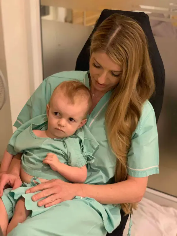
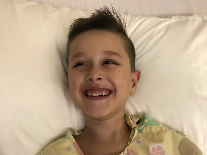
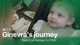
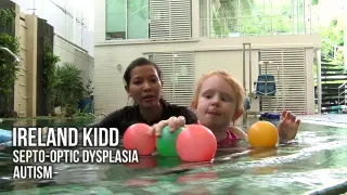
Find out more about patients previously treated with Beike stem cell protocols. The families participating in these blog posts talk about their stories and present their own view of the treatment, including thoughts regarding the daily therapies, the stem cell injection themselves as well as improvement noticed during and after treatment.
Patients and their families talking about treatment, recovery and the changes that mattered most to them.
From your first inquiry to post-treatment follow-up —
we guide you every step of the way.
Tell us about your condition. Speak with our medical advisors — no obligation.
Our doctors review your medical records and recommend a tailored protocol.
Receive specialized stem cell therapy at our partner hospital, fully supervised.
We stay in touch and monitor your progress for the months that follow.
No obligation. We’ll review your situation and respond within 48 hours.
Reviewed by the Beike medical advisory team — answering patient inquiries since 2005.