Stem Cell Treatment for Optic Nerve Atrophy (ONA): What It Involves, Who It Is For, and What the Results Show
Stem cells delivered directly to the optic nerve, not just systemically. See what visual recovery has been documented in patients with optic nerve atrophy.
62%
reported quality-of-life improvement.
59%
satisfied with the treatment outcome.
Is Stem Cell Treatment for Optic Nerve Atrophy Effective?
Are you considering Stem Cell Treatment for Optic Nerve Atrophy or Retinal Disorders?
Optic nerve atrophy and/or retinal degeneration are disabling eye disorders that have limited curative treatment options, with most only focusing on delaying disease progression and preventing further eye damage in order to ensure a better quality of life for such individuals. Stem cell treatment is being studied as a way to change that, with small studies reporting that it can improve different visual symptoms associated with optic nerve and retinal disorders (1).
Read on to see if Optic Nerve Atrophy Stem Cell Treatment might be right for you.
Is Stem Cell Therapy a Cure for Optic Nerve Atrophy?
No. Stem cell therapy does not cure optic nerve atrophy.
Nerve fibres in the optic nerve that have been permanently lost do not normally regenerate, and no treatment available today, including stem cells, has been shown to replace them (2). What the published studies and our own follow-up data describe is improvement in specific aspects of vision, such as visual acuity, light perception and visual field, in many patients, though not in all. The most likely explanation is support and neuroprotection for nerve cells that are damaged but still alive (1, 6). Clinical benefit is not guaranteed, the degree of improvement differs considerably from one person to another, and the treatment remains experimental.
What the most recent research shows
A 2024 meta-analysis of seven clinical reports found that average visual acuity improved after mesenchymal stem cell therapy in people with optic neuropathy, from 0.90 to 0.65 logMAR, while the thickness of the retinal nerve fiber layer did not change significantly. The result depends heavily on one large uncontrolled case series: when that series is removed, the improvement in acuity is no longer statistically significant (report summary, 1).
In a 2023 phase II trial in Spain, five people with a recent stroke of the optic nerve (non-arteritic ischaemic optic neuropathy) each received one injection of donor mesenchymal stem cells into the vitreous of the eye. Four of the five gained vision over 12 months. One developed a membrane on the retina that led to retinal detachment and a final vision of zero letters, another was left with a cataract, and cataract progressed in every eye that still had its natural lens. About 30% of untreated patients with this condition also improve on their own (report summary, 9). We do not inject cells into the eye.
For traumatic optic neuropathy, a 2025 review found only two human studies: one in China, where umbilical cord cells placed at the time of decompression surgery gave no significant difference in vision compared with surgery alone, and one uncontrolled series of four patients in Korea that reported improvement (report summary).
Two reviews look at future approaches. The RReSTORe consortium’s 2023 roadmap for replacing lost retinal ganglion cells reports that fewer than about 1% of transplanted cells survive in animal studies, and that reconnection to the brain has not been shown (report summary). A 2026 review of stem cell exosomes found no active clinical trials in retinal or optic nerve disease and describes safety as far from established (report summary).
So far, small and mostly uncontrolled studies report improved vision. Injections into the eye have caused harm. Regeneration of the optic nerve itself is still laboratory research. All of our summaries are listed on the optic nerve atrophy medical reports index.
Who Is a Candidate for Stem Cell Treatment for Optic Nerve Atrophy?
Timing: There is no specific timing for stem cell treatment, but we generally recommend seeking it early after diagnosis. Earlier intervention theoretically offers a better opportunity to support nerve cells that remain viable, before irreversible loss of retinal ganglion cells and their axons progresses further (2, 6).
Underlying cause: Optic nerve atrophy is the end result of many different conditions, including glaucoma, optic neuritis, ischaemic optic neuropathy, hereditary conditions such as Leber’s hereditary optic neuropathy, trauma and compressive tumours. Where the cause is still active, for example raised eye pressure or a tumour pressing on the nerve, it needs to be treated by your own specialists first, and that care continues afterwards (3, 4, 5).
Remaining vision: Stem cells are thought to support surviving nerve cells, so patients with some remaining visual function may theoretically have more to gain than those with long-standing complete loss (2, 6).
Retrobulbar injections: They are considered only for patients over 10 years old, and the treating physician decides whether they are possible.
Suitability is decided case by case. Every enquiry goes to our medical department, which reviews the patient’s medical condition through our online medical evaluation system and only then recommends a specific treatment location and protocol. We recommend speaking to our specialists before you decide, so that you know what can realistically be expected in your case.
Stem Cell Treatment for Damage to the Optic Nerve: An Encouraging Development
Stem cell therapy has become an active area of research for optic nerve damage in recent years. Stem cells are used because of their potential to protect and support damaged optic nerve cells. Stem cell therapy may improve vision and quality of life and may slow the advance of diseases that damage the optic nerve in some patients (6, 7).
Addressing Damage to Optic Nerves
Damage to the optic nerve, which is in charge of sending visual information from the eye to the brain, can lead to degeneration or injury, which can cause partial or total blindness. Damage to the optic nerve can result from a number of factors, such as (3, 4, 5, 11, 14, 15, 16):
Compressive Lesions: Growths or tumors close to the optic nerve may put pressure on it and harm it. One common example of such a lesion is a pituitary tumor.
Hereditary Conditions: Damage to the optic nerve can result from genetic disorders such as Leber’s hereditary optic neuropathy (LHON).
Trauma: A physical blow to the head or eye can cause direct damage to the optic nerve or disrupt blood flow to it.
Medication: The optic nerve may be harmed by some medications.
Lifestyle Factors: Smoking and excessive alcohol use may contribute to certain retinal or optic nerve disorders, depending on the underlying condition.
Glaucoma: Over time, increased intraocular pressure can progressively harm the optic nerve.
Optic neuropathy: Inflammation, toxins, trauma, and other factors can cause damage to the optic nerve in this condition.
Ischemic Optic Neuropathy: The optic nerve’s blood supply is diminished in this type of optic neuropathy.
Optic Neuritis: Optic neuritis is the inflammation of the optic nerve, which can be associated with multiple sclerosis (MS) and several other inflammatory or autoimmune disorders.
Identifying the Signs of Damage to the Optic Nerve
While the signs of optic nerve damage can differ, they frequently consist of (3, 4, 5, 15, 16):
Flickering or flashing lights when the eyes are moved
Persistent vision loss in one or both eyes
Either gradual or abrupt loss of vision
Diminished peripheral vision
Pain within the eye, in the eye socket, or on the face (a common sign of some conditions such as optic neuritis)
Reduced clarity of vision
Diminished ability to perceive color
Unusual reactions of the pupils to light
Variations in the optic disc’s appearance
Stem cell therapy has the potential to significantly change the optic nerve damage treatment landscape and provide hope to those afflicted by this difficult condition as research and clinical trials progress (1, 7).
Possible Improvements after Stem Cell Therapy for Optic Nerve Atrophy
Based on follow-up reports from 170 patients across 400 forms, here is the percentage who self-reported any improvement after treatment.
Symptom
% of Patients who noticed Improvement
% who noticed a Small Improvement
% who noticed a Moderate Improvement
% who noticed a Significant Improvement
Light perception
57%
37%
8%
13%
Ability to see hand movement
57%
36%
11%
11%
Pain in the eyes
53%
21%
11%
21%
Visual field
51%
35%
6%
10%
Blindness
51%
39%
6%
7%
Vision in left eye
49%
29%
11%
10%
Vision in right eye
48%
30%
9%
9%
Nystagmus (uncontrolled eye movement)
46%
14%
13%
19%
Strabismus (side glances)
45%
23%
8%
15%
Colour vision
45%
28%
6%
11%
Ability to see things at a close distance
42%
23%
11%
9%
Ability to see things clearly
42%
23%
11%
7%
Ability to focus eyes quickly
41%
23%
10%
8%
Able to count fingers
40%
21%
8%
12%
Night vision
39%
24%
7%
7%
Droopy eye lids
38%
17%
7%
14%
Ability to keep eyes focused for a long time
38%
17%
12%
9%
Ability to see things at a far distance
35%
19%
9%
8%
Patients self-assess each symptom on a 5-point scale (Worse / No improvement / Small / Moderate / Significant) at follow-up checkpoints after treatment, comparing to their pre-treatment baseline. "Reported improvement" combines the small, moderate and significant buckets. Data is updated daily from our internal patient registry. As with any medical treatment, past results do not guarantee future outcomes — improvements vary from patient to patient.
*It is important to remember that as for any medical treatment, improvements cannot be guaranteed. Please contact us for more information regarding the possible improvements for a particular case.
How Stem Cell Therapy Improves Symptoms of Retinal or Optic Nerve Disorders
Stem cells are immature cells with the ability to self-renew and, depending on the type of stem cell, develop into one or more specialized cell types. They can develop into ectodermal (ex. skin and some neurological structures), mesodermal (ex. bones, cartilages, and blood cells), or endodermal cells (ex. cells of internal body organs).
Some stem cell approaches aim to replace specific retinal cell types, while approaches based on mesenchymal stem cells (MSCs) for optic nerve disease are thought to act mainly by supporting and protecting surviving nerve cells rather than directly replacing the damaged optic nerve (6, 7). Different types of stem and progenitor cells have been investigated for retinal and optic nerve disorders. Stem cell therapy has provided new hope of improving the sight-related symptoms associated with retinal or optic degeneration in order to provide patients a better quality of life.
Following the testing of stem cell treatment on people with retinal and/or optic nerve atrophy, stem cells have been reported or proposed to have additional benefits including:
Supporting damaged retinal and optic nerve cells: Some regenerative approaches aim to replace particular retinal cell populations, and some stem cell based therapies aim to support the survival and function of remaining cells (2, 7).
Increasing the release of neurotrophic factors that may promote nervous cell proliferation and differentiation (ex. glia derived neurotrophic factor (GDNF) and brain derived neurotrophic factor (BDNF)). These factors can locally enhance cellular recruitment, proliferation and maturation within the damaged or affected retinal/optic nerve neurons (6, 12).
Modulating the immune system and the ongoing inflammatory process: Stem cells produce different antioxidants; thereby reducing the neurodestructive and atrophic process characterizing retinal degeneration and optic nerve atrophy (6, 7).
Preventing cell death: Through releasing substances that can inhibit the process of apoptosis, or programmed cell death, of the damaged cells and support stressed retinal ganglion cells (6).
Benefits of Stem Cell Therapy in Optic Nerve Atrophy and Retinal Disorders
Studies testing stem cell therapy in people with retinal and/or optic nerve disorders have reported that the use of stem cell therapy has shown improvement in (1, 8):
Visual acuity
Light perception
Color perception
Depth perception
Visual field
Night vision
In one study of six patients with dominant optic atrophy, improvement rates reported reach as high as 83%, with improvement being seen in both eyes simultaneously (report summary, 8). It was an uncontrolled study in adults treated with their own bone marrow cells. The combination of injection routes depended on the study arm. Most eyes received injections behind, beside and inside the eye, one eye was treated surgically at the retina or optic nerve, and in every patient the remaining cells were given into a vein. That is a different cell product and a different route from ours, so these figures should be read alongside the more recent research summarised above.
Safety, Side Effects and Cell Quality Control
Like any medical treatment, stem cell therapy can have side effects. Its general side effects are comparable to those of a blood transfusion or a foreign-tissue transplant: allergic reactions, cell rejection, fever. For mesenchymal stem cells given into a vein, a 2020 systematic review of 55 randomised trials, with 2,696 adult patients, found no increase in death, cancer, infection or blood-clot events with the cells, although fever occurred more often (10).
The route of injection affects the risk. In the 2023 Spanish trial of cells injected into the eye, one of five patients lost vision through retinal detachment and another developed a cataract (report summary, 9). The 2024 meta-analysis states that there were no adverse events while itself describing a patient withdrawn because of retinal detachment, so side effects are not reported consistently in this field (report summary, 1).
Our protocol delivers cells intravenously and by intrathecal injection, which is performed after a lumbar puncture, a procedure with its own risks (most commonly headache and back pain afterwards). Some patients over the age of 10 may also receive retrobulbar injections, placed behind the eyeball, near the optic nerve, and not into the eye. Retrobulbar injections can cause temporary bruising or, rarely, bleeding behind the eye (retrobulbar haemorrhage), as well as other uncommon eye or nerve injuries (13). We do not inject cells into the eye itself. The treating physicians go through all of this with you before you consent.
On the cells themselves: mesenchymal stem cells and umbilical cord blood stem cells have been studied for safety, and unlike embryonic stem cells they do not cause tumour growth (study, 10).
Every batch of cells is screened, tested twice and traceable by code before it is released for use. The full process is described on our standards and certifications page.
Where Treatment Happens, and Under Which Regulations
Treatment is delivered at affiliated hospitals in Bangkok, Thailand and in Dongguan, China. It is not approved by the US Food and Drug Administration and is not offered in the United States. It is given under Thai and Chinese regulations for cell therapy, and the cells are prepared in laboratories with CNAS, GMP and ISO accreditation. The accreditation details and the certificates are on our standards and certifications page.
Our Treatment Program in Details
We have been developing and optimizing our stem cell treatment protocols with the concept that only a very comprehensive solution can allow our patients to truly benefit from stem cells.
We believe that stimulation through various therapies is necessary to enhance stem cell regenerative response.
We provide a wide variety and large quantities of stem cells in order to adapt to each patient specific condition and deliver maximized regenerative potential.
Our stem cell treatment for Optic Nerve Atrophy (ONA) consist in 6 to 8 simple and minimally invasive injections of umbilical cord derived stem cells. The stem cells are transplanted using two or three different methods: intravenous via a standard IV drip, through intrathecal injection.
Patients older than 10 years old may also receive two retrobulbar injections to better target the optic nerves.* Together, these 3 injection methods allow for increased efficacy while ensuring safety and minimum inconvenience for the patient.
*Not all patients can receive a retrobulbar injection. The acting doctor will decide if it is possible.
01
15 to 23 Days Stay
02
IV & Intrathecal Injections
03
UCBSC / UCMSC Cells
04
Daily Therapy Program
05
120-340 Million Cells
06
Nutrition Program
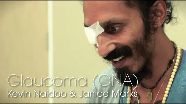
Patient Testimonial - Kevin & Janice, Glaucoma Stem Cell Treatment
Janice has suffered from visual impairment all her life, but her Glaucoma diagnosis and worsening condition coupled with a lack of treatment options was heartbreaking.
That all changed when Janice and her husband saw Kevin Naidoo’s story on local television talking about his upcoming trip for stem cell treatment.
No. Stem cell therapy is not a cure for optic nerve atrophy. Nerve fibres that have died do not normally grow back, and no current treatment replaces them (2). What studies and our own follow-up data describe is improvement in specific aspects of vision for many, but not all, patients. Improvement cannot be guaranteed and differs considerably from one patient to another.
A.
No. Stem cell therapy for optic nerve atrophy is not approved by the US Food and Drug Administration, and we do not offer it in the United States. Treatment is delivered at affiliated hospitals in Thailand and China under those countries’ regulations for cell therapy. The laboratories that prepare the cells hold CNAS, ISO and GMP accreditation, which is not the same thing as FDA approval of the treatment. More detail is on our standards and certifications page.
A.
The optic nerve is the nerve supplying the retina – the neurological part of the eye receiving different visual images to transmit them to the brain. Optic nerves, therefore, transmit visual images from the eye (retina) to the brain to be processed and analyzed. Optic nerve atrophy (ONA) is basically the loss or gradual degeneration of the optic nerve. It could occur due to hereditary causes, endocrine/metabolic causes, brain/eye tumors (ex. pituitary tumors), neurological diseases (ex. multiple sclerosis), head trauma, glaucoma, ischemia, inflammation, toxins or due to different optic neuropathies and retinal disorders – as will be discussed below (3, 4, 5). Optic nerve atrophy is usually an irreversible chronic condition. Not all optic neuropathies have a clearly reversible stage before atrophy: acute ischemic or traumatic injury, for example, can produce permanent loss rapidly.
Retinal disorders are a separate group of diseases, although severe retinal or retinal ganglion-cell disease can sometimes be associated with secondary optic-nerve changes. The most-commonly encountered retinal disorder is age-related macular degeneration (AMD), which is a major cause of visual impairment and blindness in older adults (11). Different risk factors often increase one’s risk of developing macular degeneration including smoking and cardiovascular and metabolic factors (7, 11). Although both conditions have different causes and do not necessarily follow the same pathway of degeneration and symptoms, both can cause significant and sometimes irreversible visual impairment and will therefore be discussed together in relation to regenerative and stem cell therapy.
A.
Symptoms vary according to the underlying disorder. Optic neuropathies may cause reduced visual acuity, impaired colour vision, visual-field defects or loss of contrast sensitivity, while macular retinal diseases more typically affect central vision and may cause blurred or distorted vision as the macula and fovea become involved. Symptoms may also include impaired night and/or light vision, and nystagmus, or abnormal involuntary eye movement (3, 4, 5, 11).
A.
Given that optic atrophy occurs due to nerve degeneration, its treatment options are limited similar to other neurological conditions. Once the original cause of damage causes the optic nerves to atrophy, the damage is usually irreversible and doesn’t respond to conventional treatments. Therefore, current treatment usually focuses on reducing/removing the insult damaging the retina or optic nerve prior to entering the stage of actual optic atrophy. Such treatments include:
Corticosteroids: Steroids are strong anti-inflammatory drugs that might reduce optic nerve or retinal inflammation in conditions such as optic neuritis, but they have no proven benefit in traumatic optic neuropathy and are not a general treatment for established optic nerve atrophy (5, 14).
Lifestyle modifications: These include adopting a healthy diet, exercising, smoking and alcohol cessation…etc.
Anti-VEGF drugs and other anti-angiogenic drugs: These drugs are beneficial mainly in neovascular or “wet age-related macular degeneration” and several other retinal vascular conditions which occur due to overproduction of “defective” blood vessels within the retina. Therefore suppressing the production of these blood vessels delays the retinal degeneration rate and the development of blindness (11). Several anti-VEGF therapies are now approved for this use, including ranibizumab, aflibercept, brolucizumab and faricimab. However, these drugs require prolonged, and maybe life-long, intraocular (within the eye) injections at close intervals; which might be inconvenient for many patients. Anti-VEGF treatment can preserve and often improve vision in neovascular AMD, although repeated injections are commonly required and it does not cure the underlying disease.
Photodynamic therapy (Using Verteporfin dye): This treatment is also used in wet AMD to slow the progression of the disease by targeting the abnormal blood vessels within the retina. However, this therapy doesn’t improve visual outcomes and it simply delays disease progression (11).
As you can see, treatment options are limited, and none of the mentioned treatments address the issue of retinal and/or optic nerve atrophy. Current treatments only aim to reduce the damage and/or delay disease progression. This is where stem cell therapy has been emerging in the past few years as a possible hope for the treatment of retinal degeneration and/or optic nerve atrophy after its success in improving a multitude of other neurological disorders such as cerebral palsy and autism.
A.
To date, there has not been a single study – to our knowledge – that has compared different types of stem cells, concerning safety and efficacy, particularly in patients with retinal or optic nerve disorders (1, 7). However, we can summarize different stem cell sources that have been tested in these disorders. Each form of stem cells has its own benefits and drawbacks as will be mentioned. Different stem cell sources that have been tested in ONA include:
Mesenchymal Stem Cells: These are stem cells obtained from adipose tissues, bone marrow, or umbilical cord tissues – which we actually use at Beike. These cells can be easily produced in larger numbers to accommodate higher number of patients and allow better efficacy, have better response in neurological diseases – including ONA and retinal disorders – have better immunomodulatory, neurotrophic and paracrine properties compared to other stem cells (6, 7).
Embryonic Stem Cells: Another type of stem cells includes embryonic stem cells. These cells can be differentiated into specific retinal cell types; yet they are difficult to obtain and have ethical concerns regarding their sources (7).
Induced Pluripotent Stem Cells (iPSCs): These are adult cells that have been reprogrammed into a pluripotent state and can then be differentiated toward retinal or neural cell types. Their use remains primarily experimental (7).
After carefully reviewing all of the benefits and risks of each type, we have decided to use mesenchymal umbilical cord-based stem cells that have been most extensively studied; with the least reported side effects.
In addition to the source of stem cells, there are also multiple routes of stem cell administration. Clinical trials testing stem cell therapy in ONA and retinal disorders have explored several routes, including (1, 8, 9):
Intravenous (Into the blood)
Intrathecal (Into the CSF surrounding the brain)
Retrobulbar (Behind the eye where the optic nerve resides)
Intraocular (Into the eye)
Intravitreal routes (Into the vitreous of the eye)
Sub-Tenon (Around the eye)
At Beike, we use combined intravenous and intrathecal routes; with some patients being eligible for two additional retrobulbar injections depending on different factors. We do not use intravitreal or intraocular cell injection.
A.
At Beike, we use umbilical cord stem cells for ONA and retinal disorders, both umbilical cord-related mesenchymal/tissue and blood/hematopoietic cell samples donated from healthy mothers after normal birth. As previously mentioned, this concomitant administration of both types of stem cells provides better results.
A.
There is no specific timing for stem cell treatment; but like many other neurological conditions, we generally recommend seeking stem cell therapy early after diagnosis. This is because the earlier the stem cell intervention, the easier it is to prevent further damage of the present cells and to be able to restore normal retinal or eye functioning before permanent damage takes place (2, 6). We still need to report that clinical benefit is not 100% guaranteed as is the case with any intervention, and consulting our specialists prior to undergoing the procedure is of utmost importance in order to gain more insight on the procedure and the estimated possibility of treatment success for your individual case.
A.
Of course, no treatment is without complications, and stem cell therapy is the same. However, despite its novelty, stem cell therapy has limited side effects if used properly, with comparable general side effects to those experienced with regular blood transfusion or foreign organ transplantation (ex. allergic reactions, cell rejection, or fever). Additionally, across randomised trials of mesenchymal stem cells given into a vein, no increase in death, cancer, infection or blood-clot events was found (10). Injection into the eye itself is a different matter: in a 2023 trial, one of five patients lost vision through retinal detachment (9). We do not inject cells into the eye.
A.
The following factors might influence a patient’s response to stem cell therapy, and how we at Beike Technology address each factor to ensure that we provide you with the highest efficacy using the safest procedure possible.
Dose/Number of stem cells: At Beike Technology, we administer an optimum dose of around 120-340 Million Cells (depending on the person’s weight and age) for people with different eye disorders.
Route/Method of administration: How the cells are given may affect where they travel in the body and which tissues they can reach. Combining intrathecal injection (through lumbar puncture directly within the brain’s CSF) with the traditional intravenous route may provide a better response than administering intravenous injections alone (which causes stem cells to go to other organs than the brain before reaching the brain). Other local routes, including retrobulbar and intravitreal administration, have also been investigated (1, 8, 9). Therefore, at Beike Technology, we use both intravenous and intrathecal routes concomitantly. In some selected patients, we might additionally recommend additional retrobulbar injections in order to obtain maximal efficacy; while ensuring the least possible side effects or toxicity.
Type of Stem Cells used: The type and source of stem cells may influence how they behave after treatment. We use umbilical cord-based stem cells, which have been widely studied and have shown a favorable safety profile and encouraging clinical results in a range of neurological conditions.
Timing of stem cell transplantation: As explained before, early intervention is crucial for people with optic diseases because more viable retinal ganglion cells may remain. Therefore, we recommend early intervention soon after diagnosis depending on the time one develops symptoms.
Follow-up Time: Significant benefits from stem cell therapy in patients with optic nerve and retinal diseases begin appearing weeks to months after stem cell therapy, and most people reach their full potential around 6 to 12 months after treatment – where the effects then plateau (1, 8). At Beike Technology, even after discharge, we provide you with a full follow-up program beginning as early as one month and up to one year after transplantation. You have complete access to our professional team even after you leave our center.
A.
The cause of ONA is dependent upon the type of atrophy present:
Inflammatory/demyelinating optic neuropathy, also known as optic neuritis, occurs in conditions such as multiple sclerosis and other demyelinating or inflammatory conditions (5). Patients often present with rapid loss of vision in one eye, which may be loss in part or all of the visual field.
Ischemic optic neuropathy results from occlusion of blood vessels supplying the optic nerve and can occur in conditions such as vasculitis, giant cell arteritis, granulomatosis with polyangiitis, and rheumatoid arthritis (16). Patients with ischemic optic neuropathy typically develop sudden, painless loss of vision, most often affecting the lower (inferior) half of the visual field (16).
Traumatic optic neuropathy results from direct or indirect injury to the optic nerve, often from blunt force or accidents such as motor vehicle collisions (14).
Compressive or infiltrative optic neuropathy results in the destruction of the optic nerve from locally invading tumors, infection, and autoimmune processes such as sarcoidosis (15).
Hereditary optic neuropathy: Genetic conditions including Leber hereditary optic neuropathy and dominant optic atrophy selectively damage retinal ganglion cells and their axons (3).
Glaucomatous optic neuropathy: Glaucoma causes progressive retinal ganglion-cell and optic nerve axon loss and is one of the most common causes of irreversible visual loss worldwide (4).
A.
ONA is diagnosed by extensive ophthalmological investigation which may include:
Visual field testing: visual field defects in optic neuropathies can take several patterns including central, diffuse, arcuate, and altitudinal defects (15, 16). The pattern of visual field defect is not specific to any etiology and almost any type of field defect can occur with any optic neuropathy. However, altitudinal defects are more common in ischemic optic neuropathies and central, or cecocentral defects frequently accompany toxic/nutritional and hereditary optic neuropathies.
Electrophysiological testing: Visual evoked potential (VEP) are often abnormal in optic neuropathies (5). Although VEP is not necessary for the diagnosis of optic neuropathy, it can be useful in patients with early or sub-clinical optic neuropathy who may have normal pupillary responses and no discernible optic disc changes on clinical examination.
Optical coherence tomography: OCT is a non-invasive imaging technique that uses low coherence light to penetrate tissue and a camera to analyze the reflected image. By performing circular scans around the optic nerve head, the peripapillary nerve fiber layer can be analyzed. It is widely used to evaluate structural loss and follow patients with glaucoma, optic neuritis, hereditary optic neuropathies and other optic nerve diseases (1, 3, 4, 5).
Symptoms of ONA may include blurred vision and central, peripheral (such as tunnel vision), or other localized visual-field defects (such as scotomas). These defects are diagnosed on further investigation using the aforementioned techniques.
A.
The mechanisms by which stem cells deliver their regenerative action are :
Secretion of neurotrophic factors: MSCs release certain neurotrophic growth factors including brain-derived neurotrophic factor (BDNF) which may offer neuroprotection (6).
Anti-inflammatory and immunomodulatory effects: Injection of MSC may have anti-inflammatory and neuroprotective effects that support surviving retinal ganglion cells (6, 7).
Anti-apoptotic effects: Factors secreted by MSCs may help reduce programmed cell death in stressed retinal ganglion cells (6).
Support of the local cellular environment: MSC-derived signaling molecules and extracellular vesicles are being studied for their ability to support neuronal survival and endogenous repair pathways (6, 7, 12).
Medically reviewed by
Dr. Mohammad Alzogool
Medical Director
Beike's Medical Director, Dr. Mohammad Alzogool, is a physician-researcher with a background in ophthalmology and regenerative medicine. His research contributions span ophthalmology, regenerative medicine, artificial intelligence, medical imaging, and evidence-based clinical guidelines. He brings a strong scientific and clinical perspective to the evaluation of emerging regenerative therapies.
Find out more about patients previously treated with Beike stem cell protocols. The families participating in these blog posts talk about their stories and present their own view of the treatment, including thoughts regarding the daily therapies, the stem cell injection themselves as well as improvement noticed during and after treatment.
1.
Chaibakhsh S, Azimi F, Shoae-Hassani A, et al. Evaluating the impact of mesenchymal stem cell therapy on visual acuity and retinal nerve fiber layer thickness in optic neuropathy patients: a comprehensive systematic review and meta-analysis. BMC Ophthalmology. 2024;24:316.
https://doi.org/10.1186/s12886-024-03588-2
3.
Newman NJ, Yu-Wai-Man P, Biousse V, Carelli V. Understanding the molecular basis and pathogenesis of hereditary optic neuropathies: towards improved diagnosis and management. Lancet Neurology. 2023;22(2):172-188.
https://doi.org/10.1016/S1474-4422(22)00174-0
6.
Johnson TV, DeKorver NW, Levasseur VA, et al. Identification of retinal ganglion cell neuroprotection conferred by platelet-derived growth factor through analysis of the mesenchymal stem cell secretome. Brain. 2014;137(Pt 2):503-519.
https://doi.org/10.1093/brain/awt292
7.
Van Gelder RN, Chiang MF, Dyer MA, et al. Regenerative and restorative medicine for eye disease. Nature Medicine. 2022;28(6):1149-1156.
https://doi.org/10.1038/s41591-022-01862-8
8.
Weiss JN, Levy S. Stem Cell Ophthalmology Treatment Study (SCOTS): bone marrow derived stem cells in the treatment of Dominant Optic Atrophy. Stem Cell Investigation. 2019;6:41.
https://doi.org/10.21037/sci.2019.11.01
9.
Pastor JC, Pastor-Idoate S, López-Paniagua M, et al. Intravitreal allogeneic mesenchymal stem cells: a non-randomized phase II clinical trial for acute non-arteritic optic neuropathy. Stem Cell Research & Therapy. 2023;14:261.
https://doi.org/10.1186/s13287-023-03500-7
10.
Thompson M, Mei SHJ, Wolfe D, et al. Cell therapy with intravascular administration of mesenchymal stromal cells continues to appear safe: An updated systematic review and meta-analysis. EClinicalMedicine. 2020;19:100249.
https://doi.org/10.1016/j.eclinm.2019.100249
12.
Sanie-Jahromi F, Mahmoudi A, Khalili MR, Nowroozzadeh MH. A review on the application of stem cell secretome in the protection and regeneration of retinal ganglion cells. Current Eye Research. 2022;47(11):1463-1471.
https://doi.org/10.1080/02713683.2022.2103153
14.
Blanch RJ, Joseph IJ, Cockerham K. Traumatic optic neuropathy management: a systematic review. Eye (London). 2024;38(12):2312-2318.
https://doi.org/10.1038/s41433-024-03129-7
15.
Van Stavern GP. Metabolic, hereditary, traumatic, and neoplastic optic neuropathies. Continuum (Minneapolis, Minn.). 2014;20(4 Neuro-ophthalmology):877-906.
https://doi.org/10.1212/01.CON.0000453313.37143.9b
16.
Martin-Gutierrez MP, Petzold A, Saihan Z. NAION or not NAION? A literature review of pathogenesis and differential diagnosis of anterior ischaemic optic neuropathies. Eye (London). 2024;38(3):418-425.
https://doi.org/10.1038/s41433-023-02716-4
How it works
A clear path forward
From your first inquiry to post-treatment follow-up —
we guide you every step of the way.
Stage
01
Inquiry
Tell us about your condition. Speak with our patient representatives — no obligation.
Stage
02
Evaluation
Our doctors review your medical records and recommend a tailored protocol.