Stem Cell Therapy for Spina Bifida: What It Involves, Who It Is For, and What the Results Show
Beyond surgical closure: stem cells targeting the spinal lesion to rebuild muscle strength, sensation and continence. See documented improvements in past patients.
86%
reported quality-of-life improvement.
82%
satisfied with the treatment outcome.
Is Stem Cell Treatment for Spina Bifida Effective?
Are you considering Stem Cell Treatment for Spina Bifida?
Spina bifida is a congenital neurological disorder with no current cure that can fully reverse the existing nerve damage. Most treatments focus on preventing further neurological damage and complications, managing symptoms, and preventing infections in order to improve the quality of life for such individuals. Stem cell treatment is being studied as a way to help address these limitations, with small studies reporting that it may improve some of the neurological symptoms associated with spina bifida, including movement, muscle strength, and bladder or bowel function (1, 2).
Read on to see if Spina Bifida Stem Cell Treatment might be right for you.
Is Stem Cell Therapy a Cure for Spina Bifida?
No. Stem cell therapy does not cure spina bifida.
The nerve damage in spina bifida happens before birth, and neither surgery nor stem cells can undo it completely. What the published studies and our own patient follow-up data describe is improvement in specific functions, such as bladder and bowel control, leg strength and sensation, in many patients, though not in all. Clinical benefit is not guaranteed, the degree of improvement differs considerably from one child to another, and the treatment remains experimental (1, 2).
What the most recent research shows
One of the most relevant studies to our treatment approach was published in 2019 in Stem Cell Reviews and Reports. It involved 28 children and adolescents with spina bifida who received intravenous infusions of mesenchymal stem cells derived from Wharton’s jelly. Among the 26 children with follow-up data, 21 (81%) were reported to have some improvement in their health status and quality of life. Improvements were also reported in muscle strength, movement, and bladder and bowel control, and some children gained slightly in self-care. However, the study had no control group and outcomes were rated by a single study neurologist who knew about the treatment, so it cannot confirm that these improvements were caused by stem cell treatment. The study was sponsored by the company that produced the cells (report summary, 1).
Another study of treatment after birth is a 2023 study from Vietnam of 11 children with surgically closed spina bifida and both bladder and bowel problems. Each received two intrathecal infusions of their own bone marrow mononuclear cells, six months apart. The number of children needing full help to pass stool fell from 11 to 3, and urine leakage fell from 9 children to 3, although catheter use was unchanged in 10 of the 11. The number of children able to walk independently also increased from 6 to 8. There was no control group, the study was too small to establish clinical efficacy, and only descriptive statistics were reported (report summary, 2).
The most widely reported study is the CuRe trial, published in The Lancet in 2026. Surgeons at UC Davis added a patch seeded with donor placenta-derived mesenchymal stem cells to the standard operation that closes the spine before birth. In the first six babies the patch was applied successfully, no baby had a spinal fluid leak, infection, wound breakdown or tumour detected at birth, and the hindbrain herniation reversed in all six. The paper reports results at birth only (initial safety results): it is not yet known whether adding stem cells improves movement, bladder or bowel function beyond fetal surgery alone. Follow-up continues to age six, and a larger 35-patient phase is under way. This treatment is given during fetal surgery and cannot currently be given to a child after birth in the same way (report summary, 3).
Umbilical cord cells, the type we use, have also been tested for spina bifida in experimental animal models. A 2024 study in fetal lambs published in BJOG found better movement and continence 24 hours after birth, more large nerve cells in the spinal cord and less scarring around it after treatment with a patch carrying umbilical cord mesenchymal stem cells from donor sheep, although only nine lambs were born alive (report summary, 4). Another 2024 study, using human umbilical cord cells, found more surviving large nerve cells in the spinal cord, while the difference in movement scores did not reach statistical significance (report summary, 5). A 2026 study from Germany also showed that a ready-made human UC-MSC product could be applied directly to the spinal cord during fetal spinal surgery in lambs, although only three of the seven repaired lambs survived to birth, too few to compare treated and untreated animals (report summary, 6). A 2021 systematic review of 26 preclinical studies reported encouraging results, supporting further research into stem cell treatment for spina bifida, although none of the included studies used umbilical cord cells and most had a high risk of bias (report summary, 7).
A 2026 French trial protocol (PRIUM-Cell) describes plans to test umbilical cord MSC patches during fetal surgery in 13 pregnancies, although results are not yet available (report summary, 8). A 2026 review in Seminars in Pediatric Surgery by the team running the CuRe trial describes how stem cells and patches are being combined with fetal surgery, and notes that open fetal surgery as tested in the MOMS trial remains the best-studied treatment, while stem cells before birth have so far been tested for safety in only six babies (report summary, 9). Another 2026 review of the field discusses how stem cells and biomaterials may help protect and repair damaged spinal cord tissue, but notes that, despite promising animal results, stem cell approaches in fetal repair are still far from routine clinical use (report summary, 10).
In short, small studies of stem cell treatment after birth have reported encouraging improvements in some patients, particularly in motor function, bowel and bladder control, and quality of life. The first human trial of treatment before birth has also reported promising initial safety results. However, no controlled trial has been done yet. All of our summaries are listed on the spina bifida medical reports index (1, 2, 3).
Who Is a Candidate for Stem Cell Treatment for Spina Bifida?
After surgical closure: Early surgery to close the defect remains the first and main treatment for open spina bifida. Stem cell therapy may be considered for individual evaluation afterwards, for the neurological problems that surgery does not correct. We do not offer stem cell treatment before birth; that exists only within clinical trials such as CuRe (3, 11).
Timing: Based on the trials that treated infants of different ages, we generally recommend seeking stem cell therapy early after diagnosis and surgical stabilization, during infancy or early childhood, before the window for nervous system development closes, although the optimal age for stem cell treatment has not yet been established, and in the largest study of treatment after birth age did not predict the response (1). Older children can still be assessed.
Associated conditions: Spina bifida often comes with hydrocephalus, Chiari II malformation and bladder or bowel dysfunction. The medical review looks at all of these, including whether a shunt is in place and working. Previous spinal surgery, tethered cord and the safety of intrathecal administration must also be considered (12, 13).
Medical stability: Patients with active infections, uncontrolled hydrocephalus, suspected shunt problems, or recent neurological deterioration may need further evaluation or stabilization before treatment can be considered.
What to expect: The most consistently reported gains are in bowel and bladder function and in leg strength and motor function. The degree of improvement varies, and no specific benefit can be guaranteed (1, 2).
Suitability is decided case by case. Every enquiry goes to our medical department, which reviews the patient’s medical condition through our online medical evaluation system and only then recommends a specific treatment location and protocol. We recommend speaking to our specialists before you decide, so that you know what can realistically be expected in your case.
Every patient is reviewed individually by our medical team before treatment is accepted. Treatment may be declined, postponed, or require additional medical evaluation when there is a condition that could increase the risks of stem cell administration, intravenous infusion, sedation, or lumbar puncture. Similar safety considerations have also been used as exclusion criteria in published clinical studies of cell therapy for spina bifida (2, 13).
Possible exclusion or temporary deferral criteria include:
Active infection, fever, or another acute illness at the planned time of treatment
Uncontrolled or recently active seizures, unstable hydrocephalus, suspected VP shunt malfunction, symptomatic Chiari II malformation, or another neurological condition that may increase treatment or procedural risk
Previous spinal repair or other spinal surgery, tethered cord, or significant spinal abnormalities that require further assessment before intrathecal administration (13)
An open or unhealed wound, or a skin infection, near the proposed lumbar puncture site (13)
Active cancer or a history of cancer that requires further medical assessment before treatment
Severe or uncontrolled heart, lung, liver, kidney, or other major organ dysfunction
Significant blood-clotting abnormalities or another condition that may make lumbar puncture unsafe
Severe immune deficiency, autoimmune disease requiring immunosuppressive treatment, or another significant immune-system disorder requiring individual medical assessment
A history of severe allergic reactions, particularly reactions to blood or biological products, when relevant to the planned treatment. Because latex allergy is common in spina bifida, all patients with spina bifida should be treated in a latex-free setting (14)
A medical condition that makes intravenous infusion, sedation, or intrathecal administration unsafe
Any other significant medical condition that our medical team considers to make the potential risks of treatment greater than the expected benefit
These are general safety considerations rather than absolute exclusions in every case. Some conditions may only require treatment to be postponed until the patient has recovered or become medically stable, while others may require additional investigations or modification of the treatment protocol. In particular, some patients may be suitable for intravenous administration but not for intrathecal injection. Final eligibility is determined individually after review of the patient’s medical history, current condition, medications, previous spinal surgeries, neurological status, hydrocephalus or shunt history, Chiari II malformation, relevant imaging, laboratory results and relevant medical reports (12, 13).
What Proportion of Applications Are Declined?
We do not currently publish an overall acceptance or rejection percentage. Each application is reviewed individually. Some patients are accepted immediately, while others may require additional medical information, treatment of an existing condition, a period of neurological or general medical stability, modification of the proposed protocol, or may ultimately not be considered suitable for treatment.
How Stem Cell Therapy Works to Treat Spina Bifida
Stem cells are cells that have the ability to self-renew and, depending on the type, develop into different specialized cell types. The mesenchymal stem cells used in our treatment are multipotent cells, meaning they can develop into certain specialized cell types and, importantly, release biological signals that can influence surrounding tissues (5, 7).
Therefore, injecting stem cells from a donor is thought to act mainly through the release of growth factors and other biological signals that may help protect surviving nerve cells and support tissue repair, rather than directly replacing the damaged neurons seen in spina bifida and other neural tube defects (NTDs). Stem cell therapy has provided new hope of improving the neurological symptoms associated with spina bifida, in order to provide patients a better quality of life (5, 7).
Research on stem cells in spina bifida suggests that they may offer several additional benefits beyond direct tissue replacement, including (5, 7):
Supporting and protecting damaged neurons: Rather than directly replacing spinal cord neurons, MSCs may help protect surviving nerve cells, support their function, and create a more favorable environment for neural repair and neuroplasticity (4).
Increasing the production of neurotrophic factors that support nerve cell survival, growth and function (ex. brain-derived neurotrophic factor (BDNF) and hepatocyte growth factor (HGF)). These factors can locally support the survival and function of nerve cells within the damaged or affected spinal cord tissue.
Modulating the immune system and the ongoing inflammatory process: Stem cells release different signaling molecules that may help regulate immune responses and reduce inflammation, potentially limiting further damage to vulnerable nerve tissue.
Promoting vascular supply to the nervous system: by stimulating the generation of new blood vessels (neovascularization/angiogenesis) through the release of vascular growth factors (ex. VEGF). Better blood supply may help support nerve cell survival and tissue repair.
Preventing cell death: Through releasing substances that can inhibit the process of apoptosis, or programmed cell death, which may help protect surviving nerve cells from further damage.
Reducing scar tissue formation (fibrosis): Some animal studies have reported less scar tissue around the spinal cord following stem cell treatment, significantly so in one study and as a non-significant trend in another, which may help preserve the surrounding nerve tissue and support neurological function (4, 5).
Over the last two decades, there have been numerous “animal” trials testing the efficacy and safety of stem cell treatment in-utero (during pregnancy), with encouraging findings related to nerve cell preservation, motor function and tissue repair. However, their application in humans requires further trials to prove definite efficacy (7).
The first results of the CuRe trial, the first FDA-authorized trial of stem cell therapy given before birth for spina bifida, were published in 2026 and are summarised above. They cover initial safety findings in six babies, and the additional functional benefits are still to come. Stem cell treatment before birth is available only within clinical trials (3, 8). This is why, despite the animal results on in-utero stem cell injections, we can still only use conventional stem cell therapy after birth, where stem cells have been used extensively in different neurological disorders.
Benefits of Stem Cell Therapy in Spina Bifida
Although much of the recent research has focused on administering stem cells to fetuses during prenatal surgical repair, a number of small studies have also investigated stem cell administration after birth. These include a 2018 report involving two children, a 2019 study of 28 children receiving Wharton’s jelly MSCs, and a 2023 study of 11 children receiving bone marrow mononuclear cells (1, 2, 15).
Stem cell therapy in people with spina bifida has been associated with reported improvements in the following areas (1, 2, 15):
Bowel Control
Bladder control
Lower limb power and strength
Lower limb control and movement
Bladder and bowel sensation
Gross motor development
Functional abilities and daily activities
Quality of life
In the 2019 study, 21 of 26 children (81%) with follow-up data were reported to have some improvement in their health status and quality of life, while 10 children (38%) showed a slight improvement in self-care abilities. The 2023 study also reported improvements in bowel and bladder function, with the number of children requiring full assistance with bowel evacuation decreasing from 11 to 3, and those experiencing urinary leakage decreasing from 9 to 3. In our own clinical experience, we have also observed similar types of improvements in patients with spina bifida following stem cell treatment. However, these findings and observations do not establish that the improvements were caused by stem cell treatment, and results may vary considerably between patients (1, 2).
Possible Improvements after Stem Cell Therapy for Spina Bifida
Based on follow-up reports from 22 patients across 58 forms, here is the percentage who self-reported any improvement after treatment.
Symptom
% of Patients who noticed Improvement
% who noticed a Small Improvement
% who noticed a Moderate Improvement
% who noticed a Significant Improvement
Movement in general
100%
36%
18%
46%
Overall strength
100%
27%
46%
27%
General mental ability
100%
17%
33%
50%
Interaction with others
100%
20%
20%
60%
Hand control
100%
33%
50%
17%
Trunk control
100%
22%
33%
44%
Appetite
100%
33%
33%
33%
Energy
100%
44%
33%
22%
Limb muscle strength
95%
43%
33%
19%
Trunk muscle strength
95%
30%
25%
40%
Balance
95%
25%
45%
25%
Range of movement
90%
32%
32%
26%
Fine motor control
86%
29%
57%
0%
Crawling
83%
50%
17%
17%
Spasticity
83%
33%
42%
8%
Bladder control
80%
40%
20%
20%
Standing up
75%
30%
20%
25%
Walking
74%
26%
21%
26%
Patients self-assess each symptom on a 5-point scale (Worse / No improvement / Small / Moderate / Significant) at follow-up checkpoints after treatment, comparing to their pre-treatment baseline. "Reported improvement" combines the small, moderate and significant buckets. Data is updated daily from our internal patient registry. As with any medical treatment, past results do not guarantee future outcomes — improvements vary from patient to patient.
*It is important to remember that as for any medical treatment, improvements cannot be guaranteed. Please contact us for more information regarding the possible improvements for a particular case.
How Are Improvements Evaluated After Stem Cell Treatment?
At Beike, we believe that evaluating the patient’s progress after stem cell therapy is an important part of the treatment process. Before treatment, our medical team reviews the patient’s medical history, current neurological condition, and functional abilities to establish a baseline for comparison during follow-up.
Possible improvements are evaluated through several areas, depending on the patient’s age, condition, and symptoms:
Bladder and bowel function: Changes in urinary and bowel control, episodes of incontinence, constipation, and the need for catheterization or other bowel and bladder management (12).
Lower limb muscle strength and sensation: Changes in leg muscle power, movement control, and sensation, which may be evaluated through neurological examinations and rehabilitation assessments.
Gross motor function and mobility: Changes in sitting, standing, walking, balance, and the ability to move independently or with assistance (16).
Daily functional activities: Changes in the patient’s ability to perform daily activities, such as transferring, dressing, and other age-appropriate activities.
Quality of life: Changes in comfort, independence, participation in daily activities, and the overall quality of life reported by the patient or family (17).
At Beike, we provide a follow-up program at 1, 3, 6, and 12 months after treatment. During these assessments, we review the patient’s progress through follow-up questionnaires, family observations, and relevant medical or rehabilitation reports. Additional examinations, such as urodynamic testing or neurological assessments, may also be considered when medically indicated.
It is important to understand that improvements may vary between patients, and some patients may experience limited or no noticeable changes. Comparing the patient’s condition before and after treatment helps us monitor progress over time, although any observed improvement cannot automatically be attributed to stem cell therapy alone.
Safety, Side Effects and Cell Quality Control
Like any medical treatment, stem cell therapy can have side effects. Many of the short-term reactions reported are similar to those that can occur after an IV infusion or injection, such as fever, headache, nausea, temporary discomfort, or an allergic reaction. In the published studies of stem cell therapy in children with spina bifida, the reported adverse events were generally mild and temporary, with no severe treatment-related adverse events reported (1, 2). A 2026 review of 42 randomised trials of intravenous umbilical cord MSCs in adults found that fever was more common after treatment, with no other safety signals (18).
In the 2019 study of Wharton’s jelly MSCs in 28 children, the study neurologist saw no reactions during or shortly after the infusions, and the problems parents reported in the following days, such as fever, headache, nausea and emotional sensitivity, were mild and passed without treatment (report summary, 1). The 2023 Vietnamese study of intrathecal treatment reported no severe adverse events in 11 children and seven mild ones (headache in three, and one each of vomiting, fever, urethral pain and swelling where the bone marrow was taken). All resolved without further treatment (report summary, 2). In the CuRe trial the babies had no complications attributed to the cells, although 4 of the 6 pregnancies had early rupture of the membranes, a known risk of fetal surgery itself (report summary, 3, 11).
Our protocol delivers cells both intravenously and by intrathecal injection, which is performed after a lumbar puncture, a procedure with its own risks (most commonly headache and back pain afterwards), which the treating physicians go through with you before you consent. In children who have had spinal surgery, or who have a tethered cord or a shunt, the physicians assess individually whether and where a lumbar puncture can be done safely (13).
On the cells themselves: the same 2026 review of umbilical cord MSCs given intravenously to adults found no increase in cancer or abnormal tissue growth, although follow-up was usually short and longer studies are still needed (18).
Every batch of cells is screened, tested twice and traceable by code before it is released for use. The full process is described on our standards and certifications page.
Where Treatment Happens, and Under Which Regulations
Treatment is delivered at affiliated hospitals in Bangkok, Thailand and in Dongguan, China. It is not approved by the US Food and Drug Administration and is not offered in the United States. It is given under Thai and Chinese regulations for cell therapy, and the cells are prepared in laboratories with CNAS, GMP and ISO accreditation. The accreditation details and the certificates are on our standards and certifications page.
Our Treatment Program in Details
At Beike, we have created a comprehensive therapy program to best suit patients with spina bifida. Our treatment consists of Umbilical Cord Derived Mesenchymal Stem Cells (UCMSCs) and Umbilical Cord Blood Stem Cells (UCBSCs), with simultaneous physical therapies to support neurological rehabilitation, movement, muscle strength, and daily activities.
Umbilical cord-derived stem cells come from different sources, including umbilical cord tissue and cord blood. Umbilical cord tissue contains mesenchymal stem cells, and umbilical cord blood contains hematopoietic stem and progenitor cells. Both cell types have been investigated in regenerative medicine.
Our stem cell therapy program for spina bifida consists of 6 to 8 scheduled administrations of umbilical cord-derived stem cells. The stem cells are transplanted using two separate methods: by intravenous way using a standard IV drip system, and through intrathecal injection performed after lumbar puncture. These methods are used as part of our treatment approach. Based on our clinical experience, these two delivery methods allow for increased efficacy while ensuring safety and minimum inconvenience for the patient. Intrathecal administration also requires individual medical assessment, particularly in patients with previous spinal surgery, tethered cord, or hydrocephalus. It may not be suitable for every patient.
01
15 to 23 Days Stay
02
IV & Intrathecal Injections
03
UCBSC / UCMSC Cells
04
Daily Therapy Program
05
120-340 Million Cells
06
Nutrition Program
Patient case Study - Spina Bifida Stem Cell Treatment
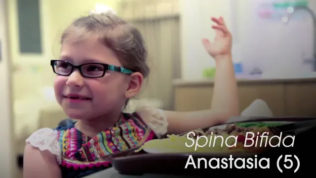
The condition is causing multiple symptoms for the 5-year-old little girl. The most pressing ones being a bladder dysfunction a lack of sensation and the lack of strength, which makes it impossible for her to crawl or walk without assistance. With the hope to improve Anastasia’s symptoms and give her more independence, her family came to Thailand to receive Stem Cell Treatment. Before going back to the USA, Anastasia’s mother and sister sat down to talk about Anastasia’s Spina Bifida the Stem Cell Treatment and the improvements they had.
No. Stem cell therapy is not a cure for spina bifida. The nerve damage begins before birth and cannot be completely undone. What studies and our own follow-up data describe is improvement in specific functions, such as bladder and bowel control, leg strength and sensation, for many, but not all, patients. Improvement cannot be guaranteed and differs considerably from one patient to another. Clinical effectiveness remains under investigation (1, 2).
A.
No. Stem cell therapy for spina bifida is not approved by the US Food and Drug Administration, and we do not offer it in the United States. Treatment is delivered at affiliated hospitals in Thailand and China under those countries’ regulations for cell therapy. The laboratories that prepare the cells hold CNAS, ISO and GMP accreditation, which is not the same thing as FDA approval of the treatment. More detail is on our standards and certifications page.
A.
Spina bifida is a congenital birth defect that occurs when the spine and the structures surrounding the spinal cord do not develop or close properly during early pregnancy. It is generally classified among Neural Tube Defects (NTDs), which involve abnormalities in the early structure that develops into the brain and spinal cord. Normally, the bones of the spine protect the spinal cord. In spina bifida, this protection may be incomplete, and in more severe forms, the spinal cord and nerves can also be affected (12).
Spina bifida is commonly described in three main forms, although other less common forms are also recognized. The severity varies depending on how the spinal cord and surrounding nerves are affected:
Spina bifida occulta: The mildest form, involving a small gap in the spine, usually without significant nerve damage or noticeable symptoms.
Meningocele: A form in which a fluid-filled sac containing the protective membranes around the spinal cord protrudes through an opening in the spine, usually with little or no nerve damage.
Myelomeningocele: The most severe form, in which part of the spinal cord and nerves are involved, potentially causing significant neurological problems.
Other, less common forms may also affect the spinal cord and nerves, even when the affected area is covered by skin. The effects of spina bifida can vary considerably from one person to another, depending on the type, location, and severity of the spinal defect (12).
A.
Symptoms of spina bifida depend on the type and severity of the condition, as well as the level of the spine affected. In general, defects higher along the spine may cause greater difficulties with leg movement and mobility. However, symptoms vary considerably from one person to another, and people with milder forms, such as spina bifida occulta, may have no noticeable symptoms.
Depending on the degree of spinal cord and nerve involvement, possible symptoms and associated problems include:
Lower limb weakness or paralysis: Difficulty moving the legs, standing, walking, or maintaining balance.
Reduced sensation: Partial or complete loss of feeling in the legs or feet, which may also increase the risk of unnoticed injuries or skin problems.
Bowel dysfunction: Constipation, difficulty controlling bowel movements, or fecal incontinence.
Orthopedic problems: Abnormalities of the feet, hips, or spine, such as clubfoot, joint deformities, or scoliosis (curvature of the spine).
Some children, particularly those with myelomeningocele, may also have associated neurological conditions such as hydrocephalus (excess fluid accumulation in the brain) or Chiari II malformation (a structural abnormality involving the lower part of the brain). These conditions may cause additional neurological problems and can affect learning, attention, coordination, or development. However, many children with spina bifida have normal intelligence, and the effects vary considerably between individuals.
Although spina bifida is present from birth, some symptoms or complications may develop or change as a child grows. New or worsening leg weakness, back pain, walking difficulties, or changes in bladder and bowel control should be medically assessed, as these may indicate complications such as a tethered spinal cord (12, 16).
A.
Spina bifida can often be detected during prenatal ultrasound examinations, particularly during the second-trimester anatomy scan, usually performed around 18 to 22 weeks of pregnancy (19, 20). Ultrasound allows doctors to examine the baby’s spine and brain, identify abnormalities, and assess the location and extent of the spinal defect. A large systematic review published in 2024 estimated that, in low-risk pregnancies, approximately 76% of spina bifida cases were detected through a single second-trimester ultrasound examination. However, detection rates can vary, and milder forms may be more difficult to identify (19).
Diagnosis may also be suspected following maternal blood screening to test for alpha-fetoprotein (AFP) in the mother’s blood. Babies create this AFP protein that commonly reaches the mother’s bloodstream, but if the level of AFP is too high it could be a sign of spina bifida. However, elevated AFP is not specific to spina bifida and requires further evaluation (10).
If further evaluation is needed, doctors may recommend a more detailed ultrasound or fetal MRI to better assess the spine, spinal cord, and associated brain abnormalities. In selected cases, amniocentesis may also be recommended to test a small sample of the fluid surrounding the baby for additional markers or genetic abnormalities. If that fluid also has high levels of AFP and acetylcholinesterase, this suggests an open defect, where the spinal cord is not covered by skin (10).
After birth, doctors can diagnose spina bifida through a physical and neurological examination. More severe forms may be visible as an opening or sac on the baby’s back, while milder forms may not be identified until later in childhood or adulthood. MRI of the spine and brain may be used when needed to confirm the diagnosis and assess the spinal cord and surrounding structures (12).
A.
Given that NTDs occur due to abnormal development of the neural tube during pregnancy, treatment options are limited similar to lots of other neurological disorders. Treatment for spina bifida depends on its type, severity, and the complications affecting each patient. Current treatment options include:
Surgical repair after birth: Surgery is the main treatment for babies born with open spina bifida (myelomeningocele). It is usually performed within the first few days after birth to close the spinal defect, protect the exposed nerves, and help prevent further damage. However, surgery cannot reverse nerve damage that has already occurred (11).
Surgical repair before birth (fetal surgery): Prenatal surgical repair has been studied in a randomized clinical trial (the MOMS trial) and is now an established option for selected cases of myelomeningocele at specialized fetal surgery centers. It is usually performed before 26 weeks of pregnancy. The trial reported clinical benefits, including a reduced need for treatment of hydrocephalus and better motor outcomes in some children compared with surgery after birth. However, fetal surgery carries significant additional risks for both mother and baby, careful patient selection remains essential, and it is not suitable for every pregnancy (11).
Treatment of neurological complications: Some patients develop hydrocephalus (excess fluid in the brain), which may require a surgically implanted shunt to drain the fluid. Other complications, such as a tethered spinal cord, may also require surgery if they cause worsening neurological symptoms or functional difficulties (12).
Bladder and bowel management: Patients with bladder or bowel control difficulties may benefit from medications, scheduled bladder emptying using a catheter, dietary adjustments, and structured bowel management programs. In more complicated cases, additional procedures or surgery may be considered. Regular monitoring of bladder and kidney function is also important to prevent long-term complications (12).
Physical and occupational therapy: Rehabilitation plays an important role in maintaining muscle strength, improving movement and coordination, preventing joint stiffness, and supporting daily activities. Depending on the patient’s needs, treatment may also include braces, walking aids, wheelchairs, or orthopedic surgery (16).
Long-term supportive care: Regular follow-up with a multidisciplinary medical team is important throughout childhood and adulthood. This may include neurological, orthopedic, urological, and rehabilitation care, together with monitoring of skin health, mobility, and other associated complications (12, 17).
While these treatments can significantly improve health and daily functioning, they do not provide a complete cure for spina bifida. Treatment is therefore focused on preventing complications, preserving existing neurological function, and helping each patient achieve the greatest possible independence.
A.
Several types of stem cells are being investigated for neurological conditions, including spina bifida. To date, no well-controlled clinical trial has directly compared the safety and effectiveness of different stem cell types specifically for spina bifida. However, we can summarize different stem cell sources that have been investigated for neurological conditions, including spina bifida. Each cell type has different biological properties, potential advantages, and limitations (1, 2, 10).
The main types being investigated include:
Mesenchymal Stem Cells (MSCs): These cells can be obtained from several sources, including adipose tissues, bone marrow, placenta, amniotic fluid, and umbilical cord tissues, which we actually use at Beike. These cells can be isolated and expanded under controlled laboratory conditions to obtain the required number of cells. MSCs are particularly interesting because of their immunomodulatory, neuroprotective and paracrine properties. Experimental studies and small clinical reports suggest possible neurological benefits in spina bifida (1, 4, 5).
Umbilical Cord Blood Stem Cells (UCBSCs): These cells are obtained from umbilical cord blood and primarily contain blood-forming stem and progenitor cells. They have been investigated for certain neurological conditions, but direct clinical evidence supporting their effectiveness in spina bifida remains limited.
Bone Marrow-Derived Mononuclear Cells (BMMNCs): These are a mixture of cells obtained from bone marrow, including stem and progenitor cells. They have been investigated for several neurological conditions. A small clinical study involving children with spina bifida reported possible improvements in bladder, bowel, and motor function, although larger controlled studies are needed to confirm these findings (2).
Embryonic Stem Cells (ESCs): Another type of stem cells includes embryonic stem cells obtained from early-stage embryos. These cells are pluripotent and can develop into many different cell types, including neural cells under specific laboratory conditions, yet they are difficult to obtain and have ethical concerns regarding their sources. Their clinical application also requires careful control of differentiation and tumorigenic risk.
Induced Pluripotent Stem Cells (iPSCs): These cells can be generated by reprogramming mature body cells, such as skin or blood cells, into a pluripotent state. They can develop into different cell types, including neurons and other nervous system cells. They provide valuable opportunities for disease modeling and regenerative research, but their clinical use in spina bifida remains at an early research stage.
Neural Stem Cells (NSCs): These are another type of stem cell being investigated for neurological conditions because they can produce neurotrophic factors and may differentiate into neural cell types under suitable conditions, but their effectiveness for treating spina bifida has not been established clinically.
After carefully reviewing all of the benefits and risks of each type, we have decided to use umbilical cord-derived mesenchymal stem cells (UC-MSCs) as our main source since they have been widely investigated in regenerative medicine and have shown encouraging safety and biological properties. This is in addition to also using umbilical cord blood cells (hematopoietic cells) concurrently to provide additional benefits from using two types of stem cells; with each contributing to neuro-regeneration through different complementary mechanisms. However, whether combining both cell types offers additional benefits for patients with spina bifida has not yet been established (18).
In addition to the source of stem cells, there are also multiple routes of stem cell administration. Clinical and experimental research has investigated different administration routes, including:
Intravenous: Administration into the bloodstream through an IV infusion.
Intrathecal: Administration into the cerebrospinal fluid (CSF) surrounding the brain and spinal cord through a lumbar puncture.
At Beike, we use combined intravenous and intrathecal routes concomitantly as part of our treatment approach when medically appropriate to achieve the best results with the least possible side effects.
A.
At Beike, we use two types of umbilical cord-derived cells for spina bifida: umbilical cord tissue-derived mesenchymal stem cells (UC-MSCs) and umbilical cord blood stem cells (UCBSCs). These cells are obtained from umbilical cords donated by healthy, medically screened mothers after childbirth. Donors undergo medical screening and infectious disease testing, while the collected cells are processed and tested under controlled laboratory conditions. As previously mentioned, we use both types of cells together to take advantage of their potentially complementary biological effects.
A.
There is no specific timing for stem cell treatment; but like many other neurological conditions and based on the available clinical studies investigating stem cell therapy in children of different ages, we generally recommend seeking an early medical evaluation after diagnosis, once the patient has undergone any necessary surgical treatment and is medically stable. This is because earlier intervention may theoretically offer opportunities to support neurological development and rehabilitation, while stem cell therapy is being investigated for its potential to protect nerve cells and reduce further neurological damage. Therefore, the most appropriate timing would be determined individually, taking into consideration the patient’s age, neurological condition, previous surgeries, and overall medical stability. We still need to report that clinical benefit is not 100% guaranteed as is the case with any intervention, and consulting our specialists prior to undergoing the procedure is of utmost importance in order to gain more insight on the procedure and the potential benefits, uncertainties, and risks for your individual case (15).
A.
Of course, no treatment is without potential complications, and stem cell therapy is no exception. However, despite its novelty, our clinical experience and published clinical studies show that stem cell therapy is generally well tolerated, with most reported side effects being mild and temporary. These may include fever, headache, nausea, or allergic reactions, similar to some reactions experienced with blood transfusions or other intravenous infusions. Additionally, in studies specifically investigating stem cell therapy in patients with spina bifida, no serious treatment-related adverse events were reported, and the observed side effects were generally mild and temporary and passed without treatment. It is also important to consider possible risks associated with the administration procedures, particularly lumbar puncture, which may cause temporary headache or back pain and, rarely, more serious complications. Although the available safety results are encouraging, larger studies and longer follow-up are still needed (1, 2, 13, 14).
A.
As with many other neurological conditions, the response to stem cell therapy may vary from one patient to another. Several patient-related and treatment-related factors may theoretically influence the results.
Factors that might affect your response to stem cell therapy for Spina Bifida include:
Severity of Spina Bifida and Remaining Neurological Function: The type of spina bifida, location of the spinal defect, extent of nerve damage, and remaining neurological function are important considerations when evaluating each patient. These factors can influence the patient’s mobility and sensation, although bladder function does not always match the level of the lesion (12, 16).
Dose/Number of stem cells: The number of stem cells administered may be an important factor when considering treatment. One preliminary study suggested a possible relationship between the total cell dose per kg of body weight and treatment response, although children who received more cells had also received more infusions (1). At Beike Technology, our treatment programmes generally involve approximately 120-340 Million Cells in total, depending on the patient’s weight, age, and individual medical condition.
Route/Method of administration: Stem cells can be administered through different routes, including intravenous infusion and intrathecal injection. These routes may provide different patterns of cell distribution and biological activity. Intrathecal administration is being investigated as a way to deliver cells closer to the central nervous system, while intravenous administration allows cells to circulate through the body. At Beike Technology, we use both routes as part of our treatment approach when medically appropriate in order to obtain the maximal efficacy. However, further studies are needed to determine whether combining these methods provides better results for patients with spina bifida than using either route alone (1, 2, 13).
Type of Stem Cells used: Different types of stem cells have different biological properties and potential therapeutic applications. Mesenchymal stem cells (MSCs) are of particular interest for neurological conditions because of their immunomodulatory and neuroprotective properties, mainly through the release of growth factors and other biological signals. At Beike, we use umbilical cord tissue mesenchymal cells as our main source of stem cells. We also use umbilical cord blood stem cells, which primarily contain hematopoietic stem and progenitor cells, to take advantage of their potentially complementary biological effects (18).
Timing of stem cell transplantation: As we previously mentioned, earlier intervention may theoretically offer opportunities to support neurological development and rehabilitation. We generally encourage families to seek an early medical evaluation after the patient has undergone any necessary surgical treatment and is medically stable.
Rehabilitation and Supportive Therapies: Physical therapy, occupational therapy, and other rehabilitation programmes play an important role in supporting movement, muscle strength, functional abilities, and independence. At Beike, we combine stem cell treatment with rehabilitation therapies to support the patient’s overall care (16).
Follow-up Time: When improvements occur, they may develop gradually rather than immediately after treatment. We generally recommend monitoring patients for possible changes over the following 3 to 6 months. At Beike Technology, even after discharge, we provide you with a full follow-up program beginning as early as one month and up to one year after transplantation. You have complete access to our professional team even after you leave our center.
It is important to understand that these factors do not guarantee a particular treatment outcome. Each patient is evaluated individually, and the potential benefits, limitations, and risks are discussed before treatment.
Medically reviewed by
Dr. Mohammad Alzogool
Medical Director
Beike's Medical Director, Dr. Mohammad Alzogool, is a physician-researcher with a background in ophthalmology and regenerative medicine. His research contributions span ophthalmology, regenerative medicine, artificial intelligence, medical imaging, and evidence-based clinical guidelines. He brings a strong scientific and clinical perspective to the evaluation of emerging regenerative therapies.
Find out more about patients previously treated with Beike stem cell protocols. The families participating in these blog posts talk about their stories and present their own view of the treatment, including thoughts regarding the daily therapies, the stem cell injection themselves as well as improvement noticed during and after treatment.
1.
Boruczkowski D, Zdolińska-Malinowska I. A retrospective analysis of safety and efficacy of Wharton's jelly stem cell administration in children with spina bifida. Stem Cell Reviews and Reports. 2019;15:717-729.
https://doi.org/10.1007/s12015-019-09902-6
2.
Nguyen LT, Le HT, Nguyen KT, Bui HT, Nguyen APT, Ngo DV, Hoang DM, Ngo MD. Outcomes of autologous bone marrow mononuclear cell administration in the treatment of neurologic sequelae in children with spina bifida. Stem Cell Research & Therapy. 2023;14:115.
https://doi.org/10.1186/s13287-023-03349-w
3.
Farmer DL, et al. Feasibility and safety of cellular therapy for in-utero repair of myelomeningocele (CuRe Trial): a first-in-human, phase 1, single-arm study. The Lancet. 2026;407:867-875.
https://doi.org/10.1016/S0140-6736(25)02466-3
4.
Athiel Y, et al. Allogenic umbilical cord-derived mesenchymal stromal cells improve motor function in prenatal surgical repair of myelomeningocele: an ovine model study. BJOG. 2024;131(6):759-767.
https://doi.org/10.1111/1471-0528.17624
5.
Athiel Y, et al. Safety and efficacy of human umbilical cord-derived mesenchymal stromal cells in fetal ovine myelomeningocele repair. Stem Cell Research & Therapy. 2024;15:444.
https://doi.org/10.1186/s13287-024-03991-y
6.
Bamberg C, et al. In vivo therapy for prenatal repair of the ovine fetal myelomeningocele model using human umbilical cord-derived mesenchymal stem cells: proof of principle. Journal of Perinatal Medicine. 2026;54(5):881-888.
https://doi.org/10.1515/jpm-2025-0577
7.
Kunpalin Y, et al. Preclinical stem cell therapy in fetuses with myelomeningocele: a systematic review and meta-analysis. Prenatal Diagnosis. 2021;41:283-300.
https://doi.org/10.1002/pd.5887
8.
Guilbaud L, et al. PRIUM-Cell : programme de réparation in utero des myéloméningocèles utilisant des cellules stromales mésenchymateuses [In utero myelomeningocele repair programme using mesenchymal stromal cells]. Gynécologie Obstétrique Fertilité & Sénologie. 2026;54(4):228-233.
https://doi.org/10.1016/j.gofs.2025.10.020
9.
Reynolds E, Rosas D, Wang A, Farmer DL. Fetal myelomeningocele repair: a narrative review of the historical perspective, emerging regenerative therapies, and future directions. Seminars in Pediatric Surgery. 2026;44:151680.
https://doi.org/10.1016/j.sempedsurg.2026.151680
10.
Evangelista A, Ruccolo L, Friuli V, et al. Advances in fetal repair of spina bifida integrating prenatal surgery, stem cells, and biomaterials. Biomedicines. 2026;14:136.
https://doi.org/10.3390/biomedicines14010136
11.
Adzick NS, et al. A randomized trial of prenatal versus postnatal repair of myelomeningocele. New England Journal of Medicine. 2011;364:993-1004.
https://doi.org/10.1056/NEJMoa1014379
12.
Radmayr C, et al. EAU-ESPU-ERN eUROGEN-ERN ITHACA-ERN ERKNet-IFSBH guidelines on spinal dysraphism in children and adolescents: summary of the guideline. European Urology Open Science. 2026;88:39-53.
https://doi.org/10.1016/j.euros.2026.04.002
13.
Engelborghs S, et al. Consensus guidelines for lumbar puncture in patients with neurological diseases. Alzheimer's & Dementia: Diagnosis, Assessment & Disease Monitoring. 2017;8:111-126.
https://doi.org/10.1016/j.dadm.2017.04.007
15.
Liem NT, Chinh VD, Thinh NT, Minh ND, Duc HM. Improved bowel function in patients with spina bifida after bone marrow-derived mononuclear cell transplantation: a report of 2 cases. American Journal of Case Reports. 2018;19:1010-1018.
https://doi.org/10.12659/AJCR.909801
18.
Hum C, Poliwoda J, Lalu M, et al. Safety of intravascular administration of umbilical-cord-derived mesenchymal stromal cells: an updated systematic review and meta-analysis. Stem Cells Translational Medicine. 2026;15(6).
https://doi.org/10.1093/stcltm/szag029
19.
Buijtendijk MFJ, et al. Diagnostic accuracy of ultrasound screening for fetal structural abnormalities during the first and second trimester of pregnancy in low-risk and unselected populations. Cochrane Database of Systematic Reviews. 2024;5:CD014715.
https://doi.org/10.1002/14651858.CD014715.pub2
20.
Salomon LJ, et al. ISUOG Practice Guidelines (updated): performance of the routine mid-trimester fetal ultrasound scan. Ultrasound in Obstetrics & Gynecology. 2022;59(6):840-856.
https://doi.org/10.1002/uog.24888
How it works
A clear path forward
From your first inquiry to post-treatment follow-up —
we guide you every step of the way.
Stage
01
Inquiry
Tell us about your condition. Speak with our patient representatives — no obligation.
Stage
02
Evaluation
Our doctors review your medical records and recommend a tailored protocol.